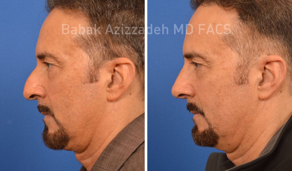
All About Rhinoplasty Austin Tx
Table of ContentsGetting My Rhinoplasty Surgeon Austin To WorkUnknown Facts About Rhinoplasty Austin Tx8 Simple Techniques For Austin Rhinopasty Surgeon
Based upon the available area of nasal skin, the surgeon chooses the place for the bilobed flap, and orients the pedicle. If the flaw remains in the lateral aspect of the nose, the pedicle is based medially. If the defect is at the nasal idea, or at the nasal dorsum, the pedicle is based laterally.
The external semi-circle defines the necessary length of the 2 lobes of the skin flap. The inner semi-circle bisects the center of the original injury, and continues throughout the donor skin, developing limitation measure of the pedicle typical to the two lobes of the flap. The cosmetic surgeon then draws 2 lines from the pinnacle of the injury; the very first line drawn is at an angle of 45 degrees from the long axis of the wound, and the second line drawn is at a 90-degree angle from the axis of the injury.
The delineation of each of the 2 lobes of the flap starts and ends at the inner semi-circle, and reaches the outer semi-circle, to the point where it intersects its central axis. The width of the very first lobe is approximately 2 mm narrower than the width of the wound; the width of the 2nd lobe is around 2 mm narrower than the width of the first lobe.
The injury is deepened, down to the nasal skeleton, to accommodate the tissue density of the bilobed flap (rhinoplasty surgeon austin). Technically, cutting the injury, expanding it, is preferable, and more secure, than trimming (thinning) the flap to fit the injury. Undermining the donor website for the 2nd lobe permits closing it mainly; it likewise eliminates excess-skin "dog-ears" at the donor site.
II. Nasolabial flap In the 19th century, the surgical techniques of J.F. Dieffenbach (17921847) popularized the nasolabial flap for nasal reconstruction, for which it remains a fundamental nose surgical treatment procedure. The nasolabial flap can be either superiorly based or inferiorly based; of which the superiorly based flap is the more useful rhinoplastic application, due to the fact that it has a more versatile arc of rotation, and the donor-site scar is inconspicuous.
Rhinoplasty Surgeon Austin Can Be Fun For Everyone
The blood supply for the flap pedicle are the transverse branches of the contralateral angular artery (the facial artery terminus parallel to the nose), navigate here and by a confluence of capillary from the angular artery and from the supraorbital artery in the median canthus, (the angles formed by the meeting of the upper and lower eyelids) (rhinoplasty austin tx).
The nasolabial flap is a random flap that is emplaced with the proximal (near) part resting upon the lateral wall of the nose, and the distal (far) portion resting upon the cheek, which includes the main angular artery, therefore is perfused with retrograde arterial circulation. Surgical technique the nasolabial flap The pedicle of the internet nasolabial flap rests upon the lateral nasal wall, and is transposed an optimum of 60 degrees, in order to prevent the "bridge effect" of a flap emplaced throughout the nasofacial angle.
The shape of the skin flap is cut from the wound template produced by the surgeon. A cut is made to the flap (without an anaesthetic injection of epinephrine), which then is raised and oriented, in an inferior-to-superior instructions, between the subcutaneous fat and the muscle fascia. The cutting continues up until the skin flap can be freely shifted upon the nasal problem.
The flap then is bent back (shown), and can be thinned (cut) under loupe zoom; however, a nasolabial flap can not be thinned as easily as an axial skin-flap. austin rhinopasty surgeon. After the nasolabial flap has been emplaced, the flap donor-site wound is sutured closed. For an injury of the lateral nasal wall that is less than 15 mm large, the flap donor-site can be closed primarily, with sutures.
Such threats are avoided by advancing (moving) the skin of the cheek towards the nasofacial junction, where it is sutured to the deep tissues. Moreover, a narrow injury, less than 1 mm wide can be enabled to heal by secondary intent (autonomous re-epithelialisation). III. The paramedian forehead flap The paramedian forehead flap is the premier autologous skin graft for the reconstruction of a nose, by replacing any of the visual nasal subunits, especially concerning the issues of different tissue density and skin color.
Limited length is a practical application limit of the paramedian forehead flap, particularly when the client has a low frontal hairline. In such a client, a little part of scalp skin can be consisted of to the flap, however it does have a different skin texture and does continue growing hair; such mismatching is prevented next page with the transverse emplacement of the flap along the hairline; yet that portion of the skin flap is random, therefore risks a greater occurrence of necrosis.
More About Rhinoplasty Austin Tx
Nonetheless, the 2nd phase of the nasal restoration can be performed with the client under local anaesthesia. Visually, although the flap donor-site scar heals well, it is obvious, and hence hard to conceal, specifically in males. Nose surgery: A paramedian forehead flap style. Surgical method the paramedian forehead flap The cosmetic surgeon creates the paramedian forehead flap from a custom-fabricated three-dimensional metal foil template originated from the measures of the nasal problem to be remedied.
Later on, the distal half of the flap is dissected and thinned to the subdermal plexus. The surgeon produces a metal foil template stemmed from the dimensions of the nasal wound. Using a Doppler ultrasonic scanner, the cosmetic surgeon identifies the axial pedicle of the tissue-flap (composed of the supraorbital artery and the supratrochlear artery), generally at the base, next to the median eyebrow; the point typically is between the midline and the supraorbital notch.